
DMI Classification
AI-powered screening and classification of Diabetic Macular Ischemia
An Automated Approach for the Screening and the Classification of Macular Ischemia
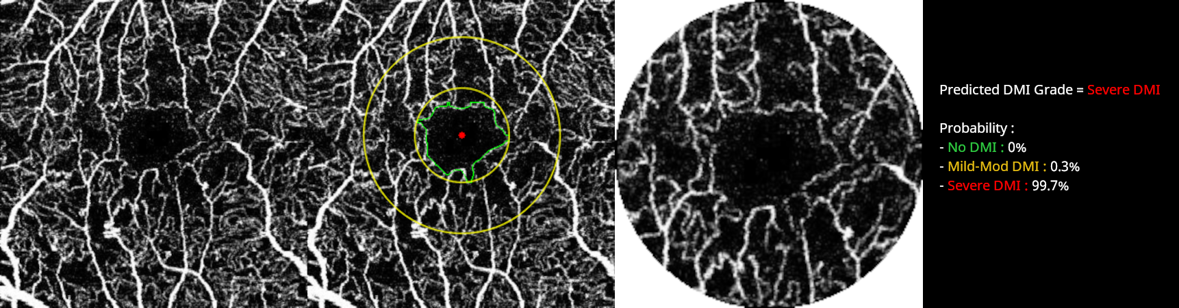
Test our deep learning model for the screening and the classification of diabetic macular ischemia in 3 × 3mm SCP Angiograms.
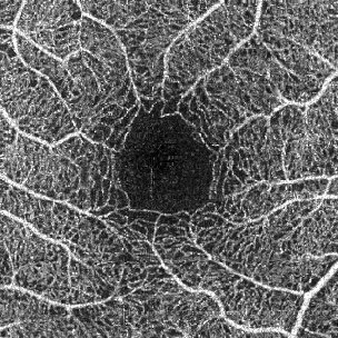
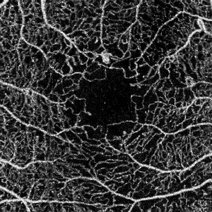
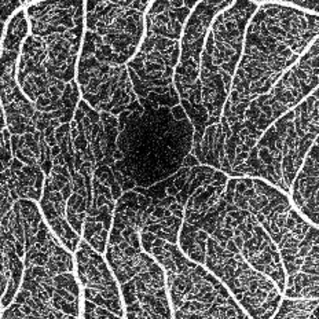
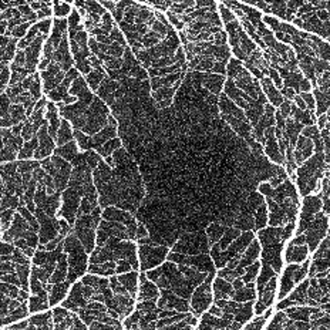
Grading of diabetic macular ischemia according to ETDRS Report No. 11 : angiograms were classified according the outline of the FAZ and perifoveal capillary loss.
| Grade | Perifoveal capillary loss | FAz Outline |
|---|---|---|
| No DMI | Absence of loss | Normal outline |
| Mild-Moderate DMI | Loss affecting less than 4 quadrants | FAZ destroyed less than 4 quadrants |
| Severe DMI | Severe loss affecting all 4 quadrants | FAZ outline completely destroyed |
MODEL STATS
Performance Metrics (%)
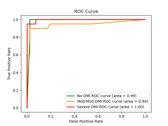
ROC Curves
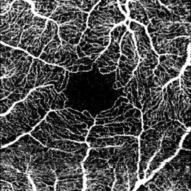
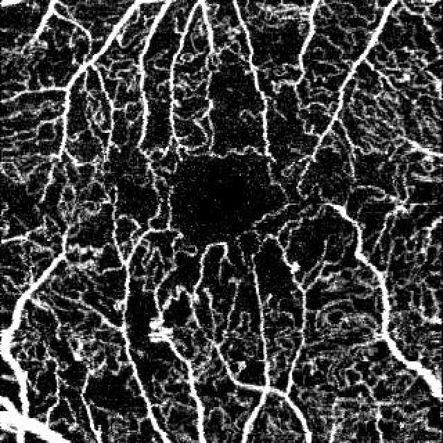
Test the model on these SCP Angiograms
SCP Angiograms of healthy and diabetic eyes from several OCT-A devices with different acquisition quality.
Or Upload your SCP Angiogram
Requirement : a raw 3 × 3mm enface OCTA image segmented to the superficial capillary plexus