FAZ Segmentation

Foveal Avascular Zone Analysis with AI Segmentation

Enhancing Foveal Avascular Zone Analysis with AI Segmentation

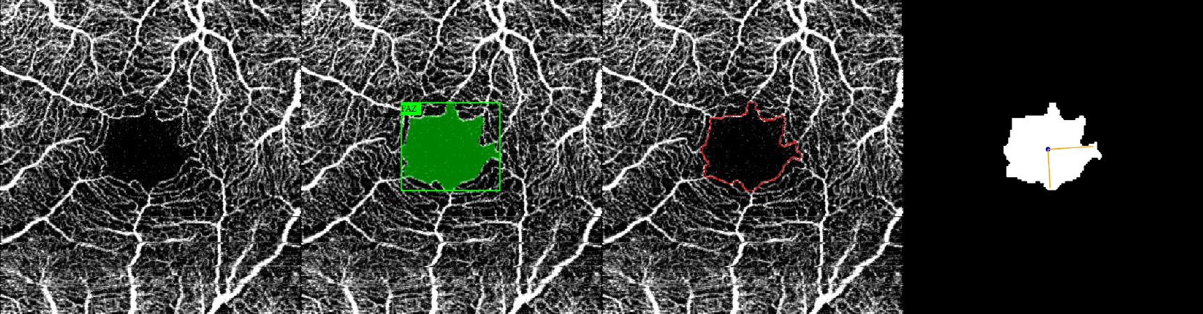
Test our deep learning model for superficial foveal avascular zone (sFAZ) segmentation in 3 × 3mm en face optical coherence tomography angiography (OCTA). Automated measurements of sFAZ biomarkers in both healthy subjects and diabetic eyes will be generated based on AI-powered sFAZ delineation.

BACKGROUND

MODEL STATS

Eye Dataset

Performance Metrics (%)

OCT-A Devices

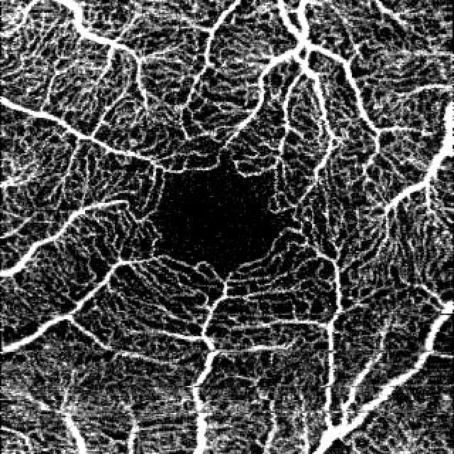
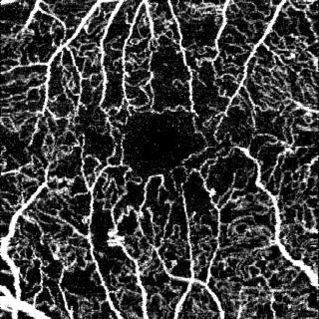
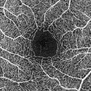
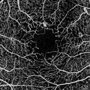
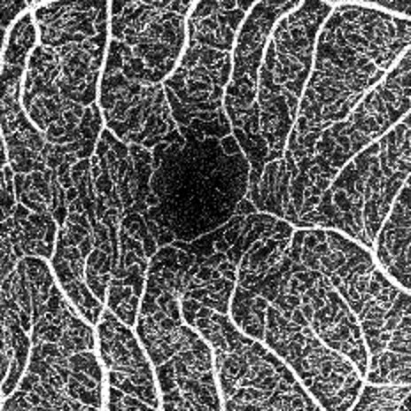
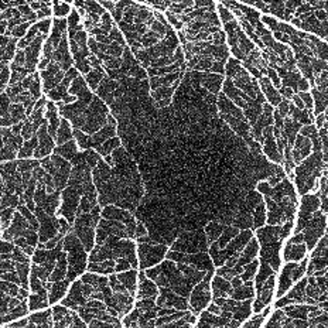
Test the model on these SCP Angiograms

SCP Angiograms of healthy and diabetic eyes from several OCT-A devices with different acquisition quality.

Good Quality Angiogram


SCP Angiogram of a Healthy Eye
Size : 3 × 3-mm = 640 × 640-pixels
Device : DRI OCT Triton-Topcon

Good Quality Angiogram


SCP Angiogram of a Diabetic Eye
Size : 3 × 3-mm = 640 × 640-pixels
Device : DRI OCT Triton - Topcon

Good Quality Angiogram


SCP Angiogram of a Healthy Eye
Size : 3 × 3-mm = 304 × 304-pixels
Device : OCT Avanti XR - Optovue

Good Quality Angiogram


SCP Angiogram of a Diabetic Eye
Size : 3 × 3-mm = 304 × 304-pixels
Device : OCT Avanti XR - Optovue

Good Quality Angiogram


SCP Angiogram of a Healthy Eye
Size : 3 × 3-mm = 464 × 464-pixels
Device : XEPHILIO OCT-A1 - Canon

Good Quality Angiogram


SCP Angiogram of a Diabetic Eye
Size : 3 × 3-mm = 464 × 464-pixels
Device : XEPHILIO OCT-A1 - Canon

Or Upload your SCP Angiogram

Requirement : a raw 3 × 3mm enface OCTA image segmented to the superficial capillary plexus

Results